Home

Salon țeavă coniac bright field live cell imaging microscope noroi recuperare A afecta

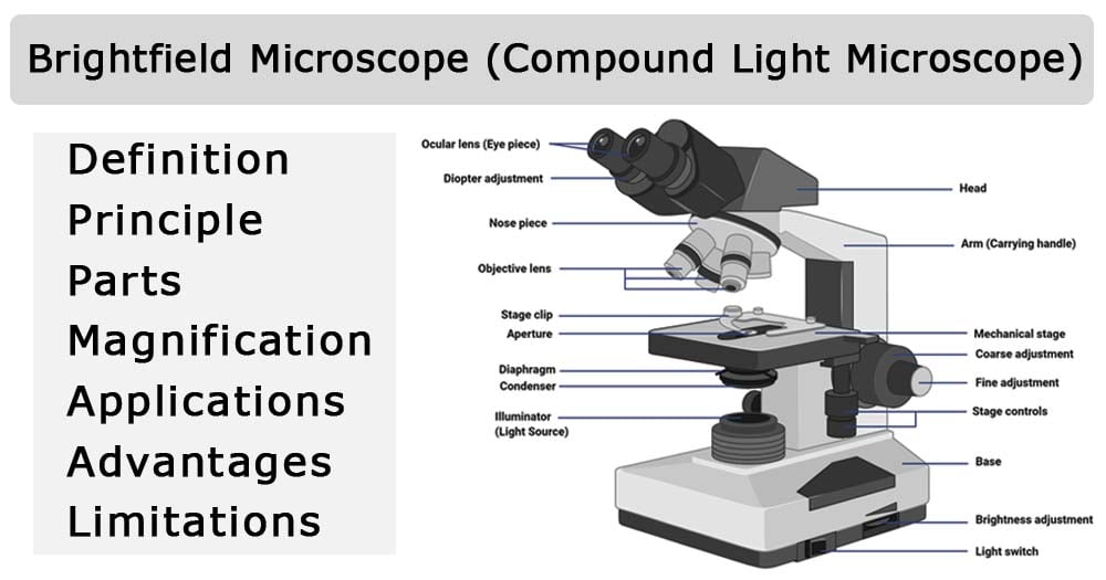
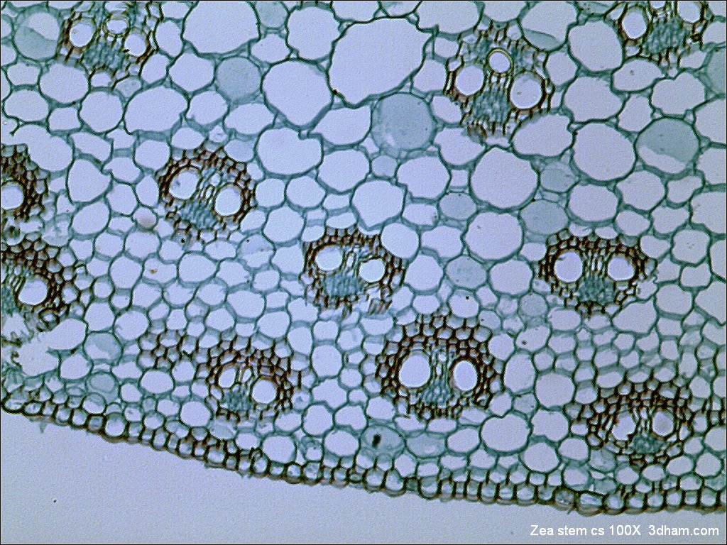
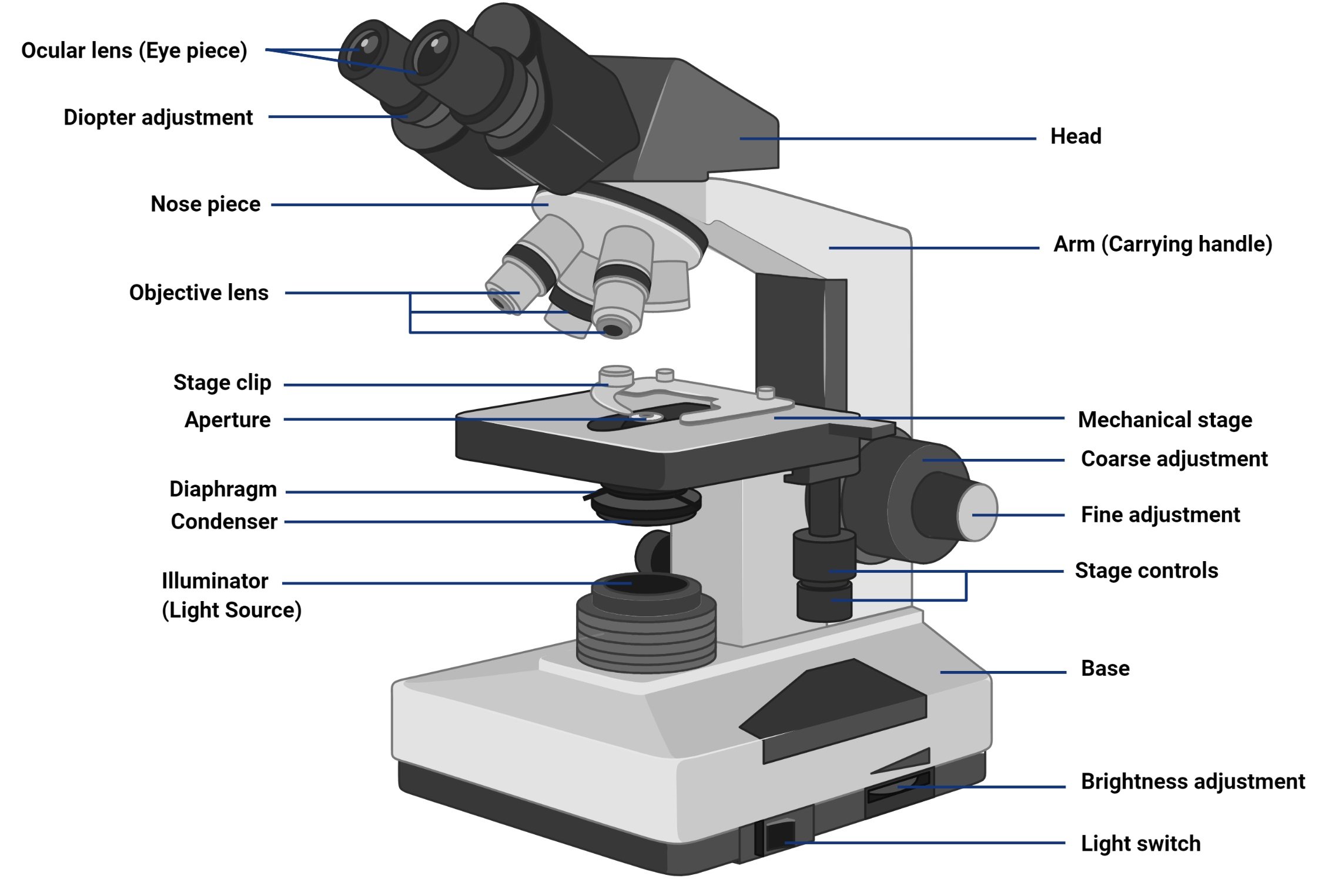
Brightfield Microscope (Compound Light Microscope)- Definition, Principle,  Parts
Brightfield Microscope (Compound Light Microscope)- Definition, Principle, Parts

Live Cell Imaging
Live Cell Imaging

11 From live LM to EM. (A) Bright-field light microscopy at 40 Â air... |  Download Scientific Diagram
11 From live LM to EM. (A) Bright-field light microscopy at 40 Â air... | Download Scientific Diagram

High-Content Screening Goes Beyond Fluorescent Pictures | Features | Aug  2016 | BioPhotonics
High-Content Screening Goes Beyond Fluorescent Pictures | Features | Aug 2016 | BioPhotonics

Cell growth and imaging quality (a) Bright field images of one imaging... |  Download Scientific Diagram
Cell growth and imaging quality (a) Bright field images of one imaging... | Download Scientific Diagram

A portable low-cost long-term live-cell imaging platform for biomedical  research and education - ScienceDirect
A portable low-cost long-term live-cell imaging platform for biomedical research and education - ScienceDirect
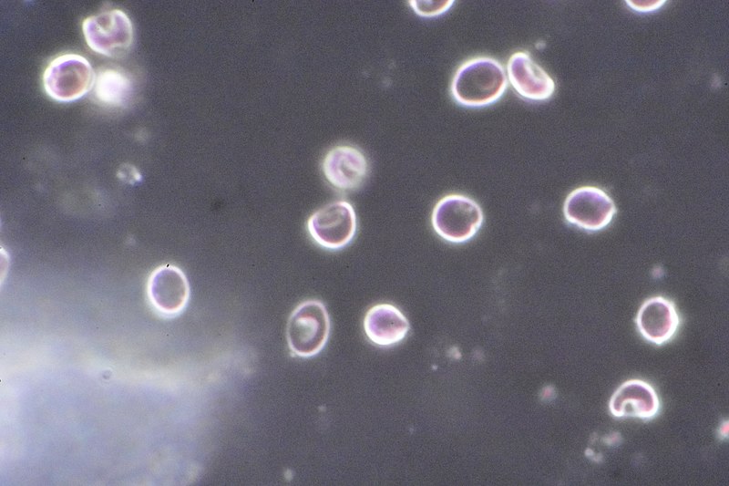
Darkfield And Phase Contrast Microscopes

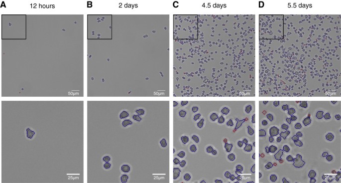
An automatic method for robust and fast cell detection in bright field  images from high-throughput microscopy | BMC Bioinformatics | Full Text
An automatic method for robust and fast cell detection in bright field images from high-throughput microscopy | BMC Bioinformatics | Full Text

Example of brightfield images of non-adherent (left) and adherent... |  Download Scientific Diagram
Example of brightfield images of non-adherent (left) and adherent... | Download Scientific Diagram

Automated Bright Field Segmentation of Cells and Vacuoles Using Image  Processing Technique - Chiang - 2018 - Cytometry Part A - Wiley Online  Library
Automated Bright Field Segmentation of Cells and Vacuoles Using Image Processing Technique - Chiang - 2018 - Cytometry Part A - Wiley Online Library
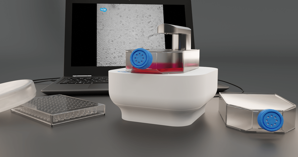
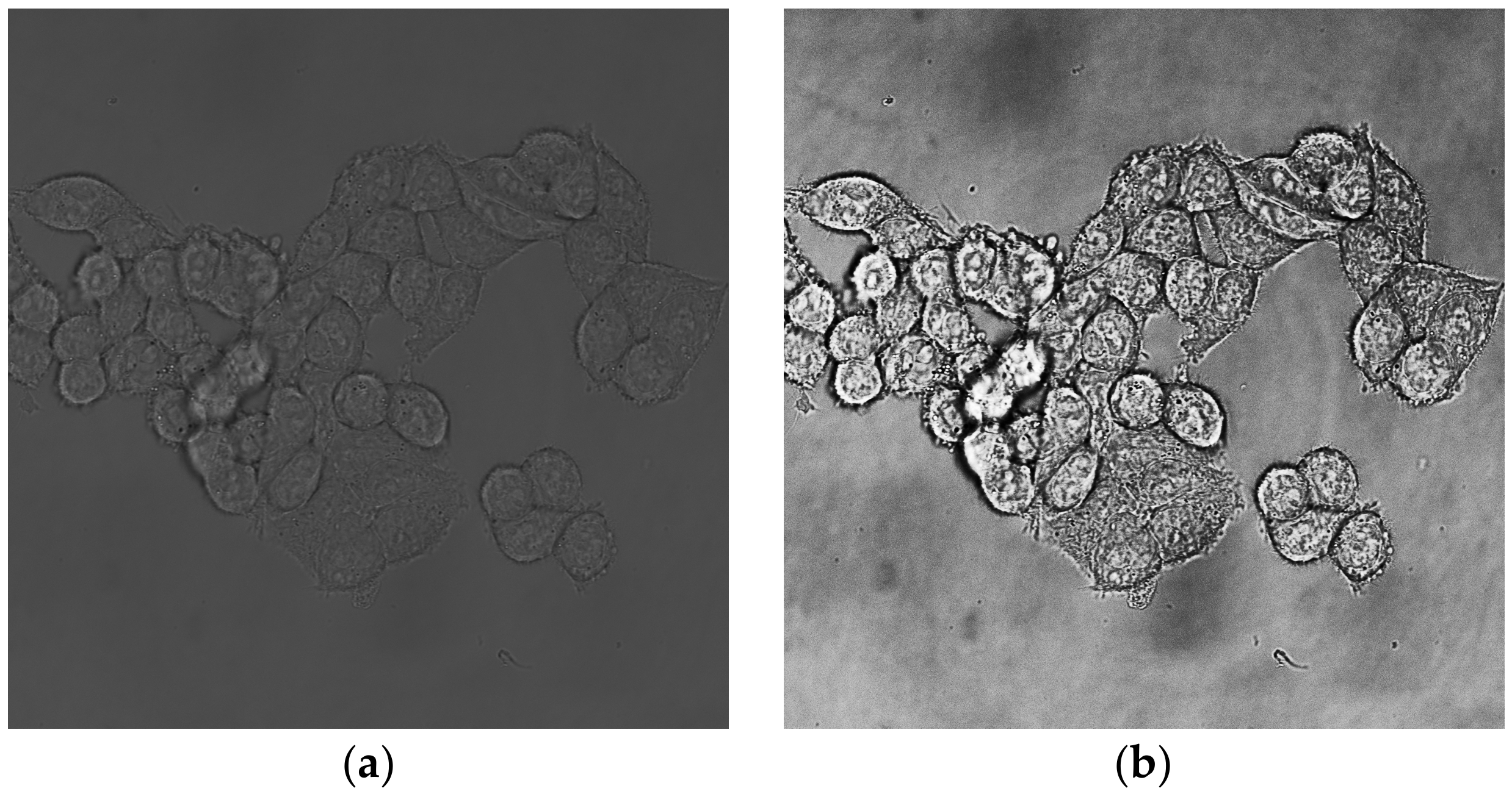
CytoSMART introduces new, label-free live-cell microscopy solutions with  high image quality
CytoSMART introduces new, label-free live-cell microscopy solutions with high image quality

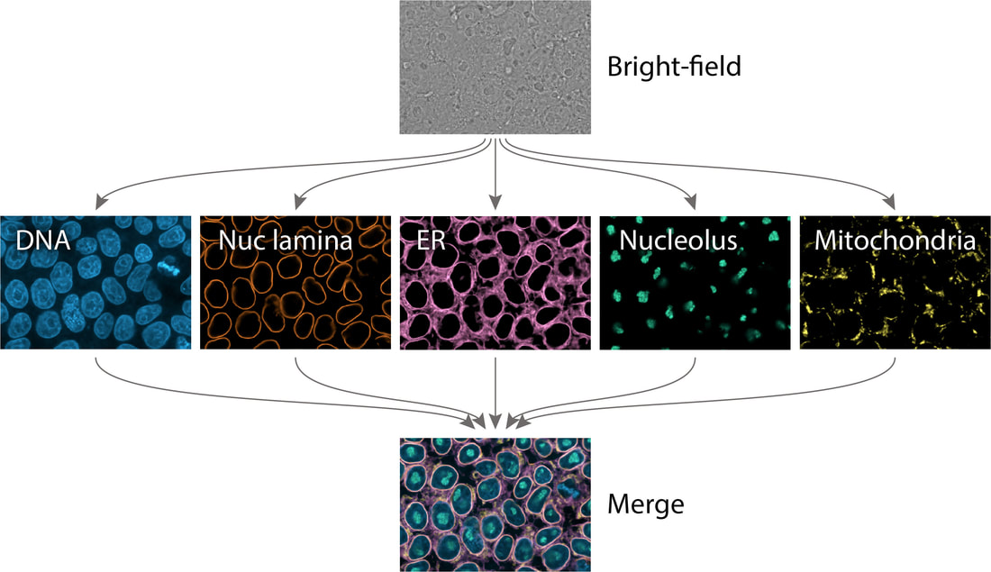
Extracting multiple cellular structures from single bright-field microscopy  images - ALLEN CELL EXPLORER
Extracting multiple cellular structures from single bright-field microscopy images - ALLEN CELL EXPLORER

Bright field (left) and fluorescence (right) images of the HEK cells... |  Download Scientific Diagram
Bright field (left) and fluorescence (right) images of the HEK cells... | Download Scientific Diagram

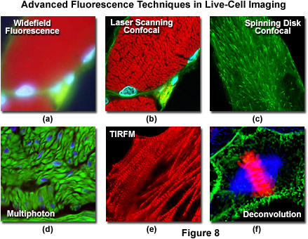
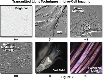
ZEISS Microscopy Online Campus | Live-Cell Imaging | Microscopy Techniques

Brightfield Microscope (Compound Light Microscope)- Definition, Principle,  Parts
Brightfield Microscope (Compound Light Microscope)- Definition, Principle, Parts

Counting unstained, confluent cells by modified bright-field microscopy |  BioTechniques
Counting unstained, confluent cells by modified bright-field microscopy | BioTechniques

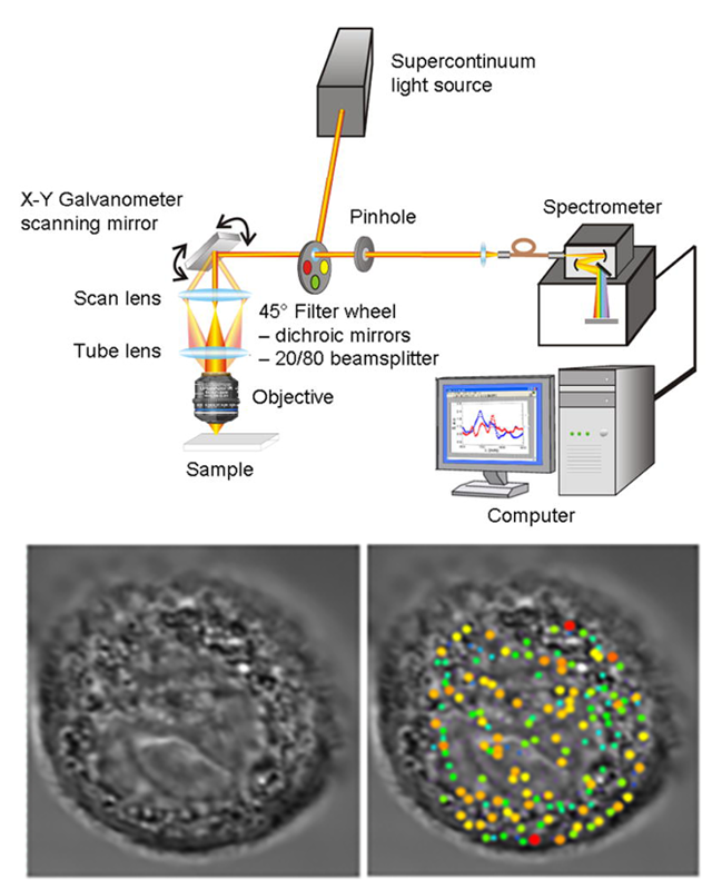
Research - Center for Advanced Biomedical Imaging and Photonics | HMS -  BIDMC
Research - Center for Advanced Biomedical Imaging and Photonics | HMS - BIDMC

ZEISS Microscopy Online Campus | Live-Cell Imaging | Microscopy Techniques

Microscopy – Membrane Enzymology

Bright-field microscopy - Wikipedia

Dark-field microscopy - Wikipedia

Bright Field Microscopy - an overview | ScienceDirect Topics

Microscopy: Dark Field, Phase Contrast, Polarization and DIC (Edward  Salmon) - YouTube
Microscopy: Dark Field, Phase Contrast, Polarization and DIC (Edward Salmon) - YouTube

MPs | Free Full-Text | Segmentation of Total Cell Area in Brightfield  Microscopy Images
MPs | Free Full-Text | Segmentation of Total Cell Area in Brightfield Microscopy Images

Live cell imaging of the HCT116 cells following treatment with 3a, 3c and  3d using bright field optical microscopy (BF) and fluorescence microscopy  (DAPI filter).
Live cell imaging of the HCT116 cells following treatment with 3a, 3c and 3d using bright field optical microscopy (BF) and fluorescence microscopy (DAPI filter).

Automated Bright Field Segmentation of Cells and Vacuoles Using Image  Processing Technique - Chiang - 2018 - Cytometry Part A - Wiley Online  Library
Automated Bright Field Segmentation of Cells and Vacuoles Using Image Processing Technique - Chiang - 2018 - Cytometry Part A - Wiley Online Library